Types of Brain Scans
The brain, our dope brain, works by generating electrical and chemical charges through its 100 billion neurons. These charges regulate the many functions of the human body, these include automated functioning such as breathing, heart beating, and blood pressure, cyclical functions such as hunger, thirst, and sleep, and perception experiences such as emotions, thoughts, and a conscious awareness of the mind itself.
It’s a very complex and beautiful ballet of nanoscopic activity. Brain cells produce neurotransmitters, nerve fibers convey pain or move our limbs, and sense organs turn light rays or soundwaves into electrical signals. All of these attributes of the human experience have been studied in the body and only relatively recently have scientists begun to trace these qualities to specialized areas of the brain.
Thanks to modern technology, there are several ways to measure brain activity to help locate tumors, detect problem areas that caused a stroke, and visualize the many components of the human condition.
There is a plethora of research showing neurological benefits during activities of relaxation, meditation, and creative expression. To set ourselves up for a deep dive into these articles, it’s helpful to know what tools these scientists are utilizing so we can follow along during this wonderful age of self-realization.
Electroencephalography (EEG)
Electroencephalography (EEG) is a functional imaging process that records signals from the electrical activity caused by nerve cell firing. It maps brainwaves in different states of mind and helps diagnose sleep disorders, epilepsy, and brain dysfunctions. The above pictures are from when I got an EEG to see if a brain dysfunction was the cause of my restless leg syndrome. To test how my brain functioned while I slept, I was required to not sleep the night before. During the process, they asked that I remain in a calm state and allow myself to fall asleep. Another part included flashing lights that were designed to trigger epilepsy which I found very uncomfortable. Although I didn’t get cool brainwave images in my medical records, the results showed that my brain was normal. Overall, I found the EEG testing to be fascinating.
Position Emission Tomography (PET)
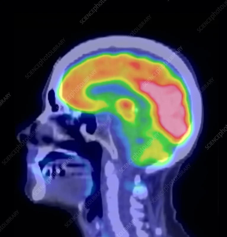
Position Emission Tomography (PET) is a functional scanning technology that measures alterations at a biological level. By injecting a specialized radioactive marker, the PET scan can detect the reactive gamma waves, pinpoints their exact location, and depict a three-dimensional image of the brain. It’s a very intricate process that’s best explained in this video.
Functional Magnetic Resonance Imaging (fMRI)
Functional Magnetic Resonance Imaging (fMRI) is a functional scanning process developed in the early 1990s that measures the change in blood flow and electrical activity in specific areas of the brain. It uses the magnetic field created by electrical activity in the brain and detects the change in O2 which changes the magnetic field. It is used to track higher oxygenated blood in a particular region and map the brain to the task performed.
Magnetic Encephalography (MEG)
Magnetic Encephalography (MEG) is a functional scanning process that traces the brain’s magnetism. It only maps the electrical activity of the brain so they are paired with MRI scans which show anatomical structure. MEG is often used to map the activities of the brain but is also used to control computer and mechanical operations by only using the mind.
Diffusion Sensor Imaging (DSI)
Diffusion Sensor Imaging (DSI) is a functional scanning process also known as diffusion MRI or Tractography. It is used to track water molecules along nerve fibers that reveal the brain’s long-distance connection. The colored spaghetti is the axons connecting neurons and shows where the information is flowing.
Computed Tomography (CT)
Computed Tomography (CT) scans reveal the anatomical features of the brain but do not resolve its structure well. This combines computer imaging with x-rays to allow for different viewing angles. It provides the structure of the brain to compare soft tissue, bone, and blood to locate tumors and blood clots. Although it’s less detailed, it’s the quickest way to see any major differences and is the first scan after the symptoms of a stroke.
Magnetic Resonance Imaging (MRI)
Magnetic Resonance Imaging (MRI) is the most common anatomical imaging tool and is used to read brain tissues in a non-invasive way. With the use of magnets and echoes of radio waves, an MRI scan can construct a detailed composition of the brain. It can show how blow flows through certain blood vessels or water in the spinal fluid distribution in the brain.
Functional Near Infrared Spectroscopy (fNIRS)
Functional Near Infrared Spectroscopy (fNIRS) is a functional scanning technique for measuring blood oxygenation in the brain. It is similar to fMRIs but instead of laying still, fNIRS can be studied during movement, even while playing an instrument. Near Infrared light can be absorbed by the blood, fat, and different levels of blood oxygenation. By shining this light, fNIRS can measure activities in different areas.
January 26, 2023


